Tissue Alterations in Oreochromis niloticus Following Chronic Exposure to Metal Complex Dark Green Azo Acid Dye and Anionic Surfactant Oil
Article Sidebar
Main Article Content
Abstract
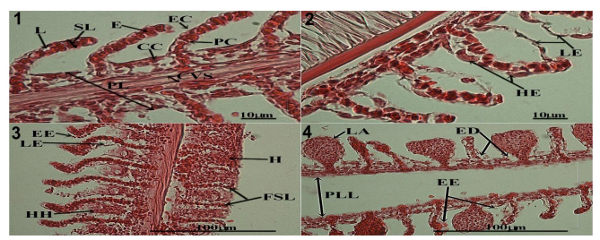
Gill, liver and kidney tissues in Oreochromis niloticus underwent histological alterations during a 90-day chronic exposure to metal complex dark green azo acid dye; anionic surfactant oil or mixtures of the two substances. Gill alterations following these chronic exposures included primary lamellae lifting, epithelial hypertrophy, secondary lamellae hyperplasia, secondary lamellae tip fusion, lamellae aneurysm and fusion, edema and blood congestion, all reflective of impaired metabolism and ion exchange. Liver alterations included cytoplasm degeneration, dilated sinusoid blood vessels, pyknotic nuclei, karyolysis, cytoplasm vacuolation and blood congestion suggesting reduced detoxification function. Kidney changes included tubule degeneration, dilation of glomeruli capillaries and Bowman’s space indicating excretory difficulties. Necrotic kidney tissue was found in fish exposed to 6 mg/Lmetal complex dark green azo acid dye. Histological examination of tissues following chronic exposures to toxic substances facilitates early diagnosis and understanding of the mechanisms by which substances impose harmful effects on organisms.
Article Details
Published articles are under the copyright of the Environment and Natural Resources Journal effective when the article is accepted for publication thus granting Environment and Natural Resources Journal all rights for the work so that both parties may be protected from the consequences of unauthorized use. Partially or totally publication of an article elsewhere is possible only after the consent from the editors.