Impact of Dual Energy Window Scatter Correction on Image Quality in Tomographic Iodine 131
Article Sidebar
Main Article Content
Abstract
Background: The major limitation factors in single photon emission computed tomography (SPECT) detectability is the presence of scattered photons within the main photopeak.
Objective: To evaluate the impact of dual-energy window (DEW) scatter correction from Xeleris processing workstation (GE Healthcare) on the image quality including image contrast, background noise, and contrast-to-noise ratio (CNR) .
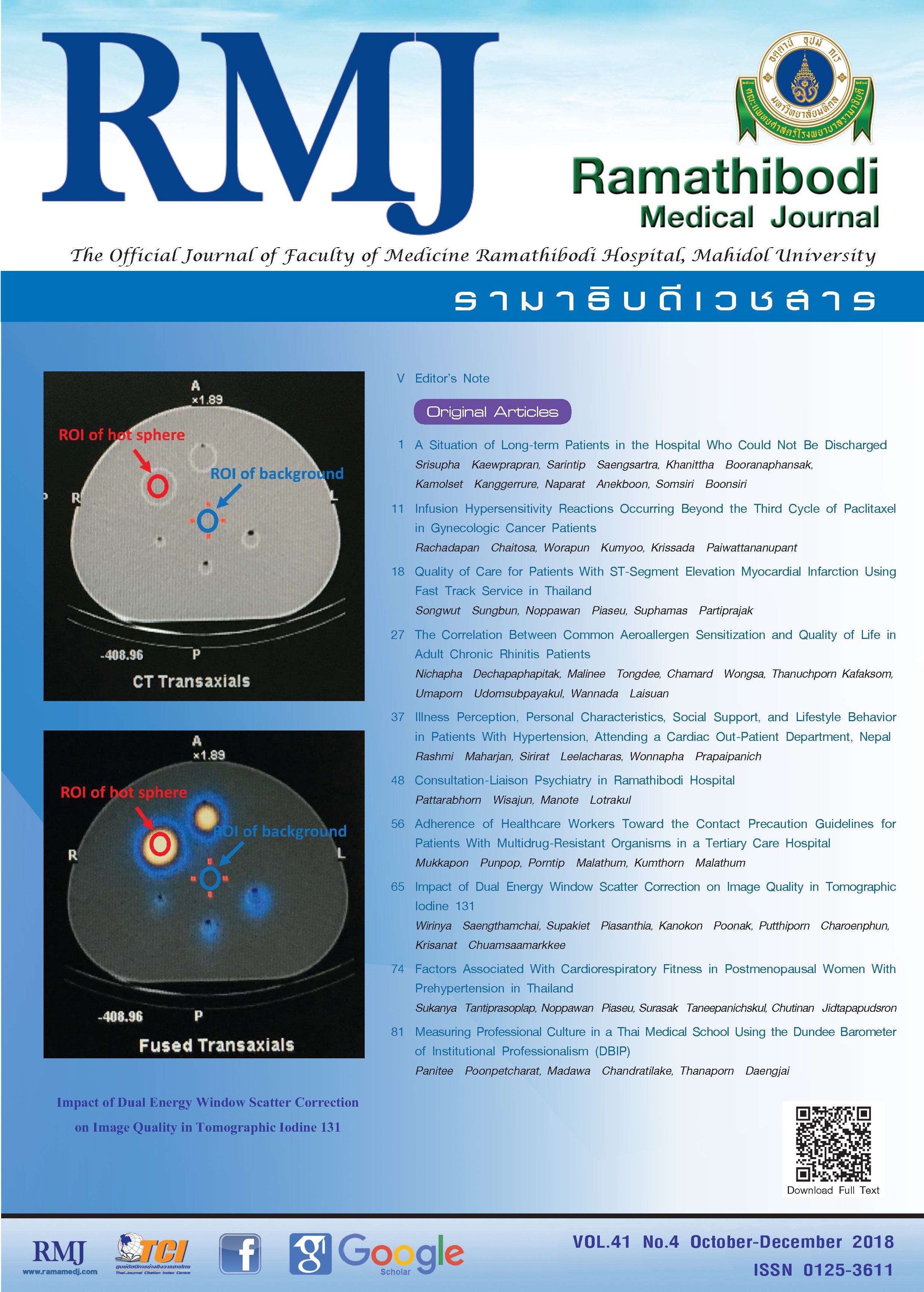
Methods: A NEMA/IEC phantom with a set of fillable spheres was used in this study. The phantom with hot lesion to warm background ratio of 10:1 were acquired with different acquisition times 20, 30, 40, and 90 seconds per frame. All projections were reconstructed with no scatter correction (NSC) and scatter correction (SC) using Xeleris vendor-supplied software.
Results: Image contrast decreased as the acquisition time increased and the larger sphere showed the greater contrast as expected. Interestingly, the contrast dropped more than half when applied SC compared with NSC. For noise, the SC resulted in an increased of image noises for all sphere sizes and acquisition times when compared with NSC. To reveal the impact of scatter on contrast and noise, the CNR was investigated, our results found that the CNR for SC was higher than NSC when acquired 30, 40, and 90 seconds per frame for small spheres (0.52 mL, 1.15 mL, and 2.57 mL), whereas in large sphere (26.52 mL and 11.49 mL), the CNRs were varied when SC or NSC applied.
Conclusions: Our results showed that the implementation of DEW scatter correction could be degraded image quality in an iodine 131 (131I) SPECT when compared with uncorrected image.
Article Details


References
2. Dewaraja YK, Ljungberg M, Green AJ, et al. MIRD pamphlet No. 24: guidelines for quantitative 131I SPECT in dosimetry applications. J Nucl Med. 2013;54(12):2182-2188. doi:10.2967/jnumed.113.122390.
3. Asl M, Sadremomtaz A, Bitarafan-Rajabi A. Evaluation of six scatter correction methods based on spectral analysis in (99m)Tc SPECT imaging using SIMIND Monte Carlo simulation. J Med Phys. 2013;38(4):189-197. doi:10.4103/0971-6203.121197.
4. Knoll P, Rahmim A, Gultekin S, et al. Improved scatter correction with factor analysis for planar and SPECT imaging. Rev Sci Instrum. 2017;88(9):094303. doi:10.1063/1.5001024.
5. Sadrmomtaz A, Kakasoltani S. Scatter correction with dual energy window method in SPECT and investigation the effect of scatter correction on image contrast value in reconstructed data. World Applied Programming. 2011;1(3)150-154.
6. van Gils CA, Beijst C, van Rooij R, de Jong HW. Impact of reconstruction parameters on quantitative I-131 SPECT. Phys Med Biol. 2016;61(14):5166-5182. doi:10.1088/0031-9155/61/14/5166.
7. Erdi YE. Limits of tumor detectability in nuclear medicine and PET. Mol Imaging Radionucl Ther. 2012;21(1):23-28. doi:10.4274/Mirt.138.
8. Smith MF, Jaszczak RJ. Generalized dual-energy-window scatter compensation in spatially varying media for SPECT. Phys Med Biol. 1994;39(3):531-546.
9. Beijst C, Kist JW, Elschot M, et al. Quantitative comparison of 124I PET/CT and 131I SPECT/CT detectability. J Nucl Med. 2016;57(1):103-108. doi:10.2967/jnumed.115.162750.
10. Elschot M, Vermolen BJ, Lam MG, de Keizer B, van den Bosch MA, de Jong HW. Quantitative comparison of PET and Bremsstrahlung SPECT for imaging the in vivo yttrium-90 microsphere distribution after liver radioembolization. PLoS One. 2013;8(2):e55742. doi:10.1371/journal.pone.0055742.
11. Narita Y, Eberl S, Iida H, et al. Monte Carlo and experimental evaluation of accuracy and noise properties of two scatter correction methods for SPECT. Phys Med Biol. 1996;41(11):2481-2496.